Logo

Experience a tailored walkthrough of our solution and find out how it fits your workflow perfectly.

Contact Us

Get early access

Stay updated with our latest news and offers.

Follow Us

Ophthalmology EHR with ophthalmic imaging, visual acuity tracking, and AI clinical documentation

Ophthalmology EHR with ophthalmic imaging, visual acuity tracking, and AI clinical documentation

Digital Patient Chart supports ophthalmology practices with ophthalmic imaging through the built-in DICOM viewer, detailed eye exam templates, and longitudinal tracking of visual acuity, intraocular pressure, and disease progression. OCT scans, fundus photos, visual fields, and fluorescein angiography attach directly to the patient record. The AI scheduling engine coordinates providers, diagnostic equipment, and procedure rooms so patients move through refraction, diagnostic testing, and examination without equipment conflicts.

  • Ophthalmic imaging inside the patient record

    OCT scans, fundus photography, visual field maps, and fluorescein angiography display through the built-in DICOM viewer with local PACS storage. Images attach to the visit record and compare against prior studies within the same chart.

  • Detailed eye exam SOAP templates

    Build SOAP fields for visual acuity, refraction, slit lamp findings, tonometry, dilated fundus exam, and specialized testing results. The AI maps voice documentation to these fields during the encounter.

  • Visual acuity and IOP tracking across visits

    Visual acuity measurements, intraocular pressures, and visual field results track longitudinally. The provider sees trends in disease markers over time to assess treatment response for glaucoma, macular degeneration, and diabetic retinopathy.

  • Diagnostic equipment and procedure room scheduling

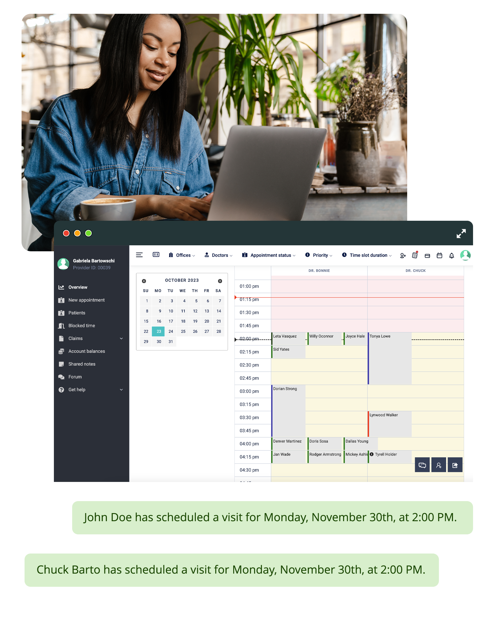
    The scheduling engine manages providers alongside OCT machines, visual field analyzers, laser equipment, and procedure rooms. Equipment reserves automatically with the appointment.

What the platform does for ophthalmology practices

Specialty-specific SOAP templates for ophthalmic exams

The form builder creates fields for visual acuity (distance, near, with correction), refraction data, slit lamp findings by structure, Goldmann or NCT tonometry, gonioscopy, dilated fundus exam per eye, and cup-to-disc ratios. The AI maps voice dictation to these structured fields. Providers document a complete eye exam without manual data entry.

DICOM viewer for ophthalmic imaging

OCT scans, fundus photos, visual field plots, fluorescein angiography, and corneal topography display through the native DICOM viewer with local PACS storage. Images attach to the specific visit record. On follow-up visits, providers compare current imaging against prior scans to track retinal thickness changes, optic nerve head progression, or corneal changes.

Longitudinal disease monitoring

For glaucoma patients: IOP readings, visual field progression, and RNFL thickness trend over time. For macular degeneration patients: OCT thickness measurements and injection counts track per eye. For diabetic retinopathy patients: visual acuity changes and retinal findings document alongside HbA1c values. Each disease tracks independently per eye.

Diagnostic equipment scheduling

OCT machines, visual field analyzers, fundus cameras, and laser units schedule as resources alongside provider appointments. When an OCT scan is ordered, the machine reserves for that time slot. Multiple ophthalmologists sharing the same equipment will not encounter double-bookings.

Per-eye documentation

Ophthalmology documentation requires per-eye specificity. The SOAP template separates findings by OD and OS with independent fields for each. The AI recognizes laterality during voice documentation and maps findings to the correct eye.

Pre-visit analysis for ophthalmic patients

Before the patient arrives, the AI reviews prior imaging, IOP trends, visual acuity changes, treatment history (injection counts, laser dates), and medication compliance per eye. The provider starts the visit knowing which eye has changed and what the treatment trajectory looks like.

Ophthalmology billing with procedure and testing codes

CPT codes for office visits, comprehensive eye exams, OCT, visual fields, fundus photography, fluorescein angiography, intravitreal injections, and laser procedures populate from documented services. Each service line carries the correct code and modifier. The invoice generates at checkout.

Structured Onboarding for Ophthalmology Practices

Ophthalmology practices rely on imaging workflows that need to integrate with clinical documentation and per-eye tracking from the start. Our onboarding team configures the DICOM viewer and local PACS, builds ophthalmic exam SOAP templates with per-eye fields, sets up diagnostic equipment scheduling, and trains providers on AI voice documentation with ophthalmic terminology so imaging-integrated exams work from the first week.

Read More
Structured Onboarding for Ophthalmology Practices

Frequently Asked Questions

Shape

Is there an EHR with a built-in viewer for OCT and retinal imaging?

Digital Patient Chart includes a native DICOM viewer with local PACS storage for OCT scans, fundus photography, visual field plots, fluorescein angiography, and corneal topography. Images attach to the visit record and compare against prior studies within the same chart.

Can the system track visual acuity and IOP across visits?

Visual acuity measurements, intraocular pressures, and visual field results track longitudinally per eye inside the patient record. Providers see trends over time to assess treatment response for glaucoma, macular degeneration, and diabetic retinopathy.

Does the EHR support per-eye documentation?

The SOAP template separates findings by OD and OS with independent fields for each eye. The AI recognizes laterality during voice documentation and maps findings to the correct eye automatically.

Can the scheduling engine manage OCT machines and visual field analyzers?

The scheduling engine manages diagnostic equipment as resources alongside provider appointments. When an OCT or visual field test is booked, the equipment reserves for that time slot. Multiple providers sharing equipment will not encounter double-bookings.

Does the AI review ophthalmic history before appointments?

Before the patient arrives, the AI reviews prior imaging, IOP trends, visual acuity changes, treatment history per eye, and medication compliance. The provider starts the visit knowing which eye has progressed and what the treatment trajectory shows.

How does billing work for ophthalmology procedures and testing?

CPT codes for eye exams, OCT, visual fields, fundus photography, angiography, injections, and laser procedures populate from documented services. Each service line carries the correct code and modifier. The invoice generates at checkout.

See ophthalmic imaging, per-eye tracking, and AI documentation in a live demo

See ophthalmic imaging, per-eye tracking, and AI documentation in a live demo

Get a Demo

We use first-party cookies to run this site and understand how patients find us. Privacy